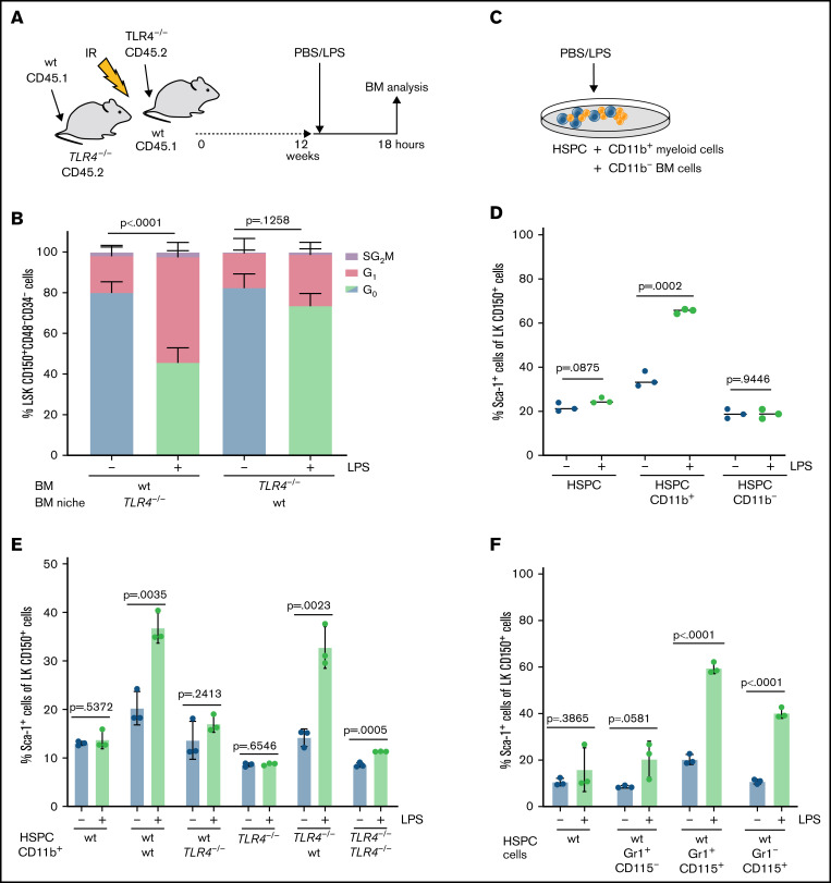
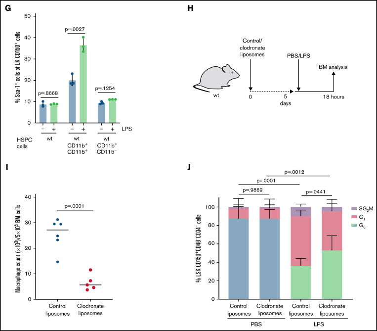
Figure 3.
Systemic LPS challenge induces HSC activation via CD115+ monocytic cells in BM. (A) Scheme indicating in vivo transplantation: BM from wt or TLR4−/− mice was transplanted (3 × 10⁶ BM cells) into irradiated TLR4−/− or wt mice. Chimeric mice were treated with PBS (control) or LPS (0.25 mg/kg, 18 hours) 12 weeks after transplantation. (B) Cell cycle analysis (icKi67-Hoechst 33342) of wt and TLR4−/− HSCs (LSK CD150+CD48-CD34-) in forward and reverse chimeric mice after PBS (control) or LPS treatment as indicated in Figure 3A (n = 6). P values refer to G0 phase and were determined by ANOVA Tukey’s post hoc test. (C) Scheme indicating in vitro culturing of sorted HSPCs (LK CD150+) with CD11b+ myeloid cells or CD11b- BM cells in presence of PBS (control) or LPS (100 ng/mL, 18 hours). (D) Relative Sca-1 expression on sorted wt HSPCs (LK CD150+) after in vitro culture for 18 hours in the presence of wt CD11b+ or CD11b- BM cells treated with PBS (control) or LPS (100 ng/mL) as indicated in Figure 3C (n = 3). P values were determined by unpaired t test. (E) Relative Sca-1 expression on sorted wt or TLR4−/− HSPCs (LK CD150+) after in vitro culture for 18 hours in presence of wt or TLR4−/− CD11b+ myeloid BM cells treated with PBS (control) or LPS (100 ng/mL) (n = 3). P values were determined by unpaired t test. (F) Relative Sca-1 expression on sorted wt HSPCs (LK CD150+) after in vitro culture for 18 hours in the presence of wt Gr1+CD115- BM cells, Gr1+CD115+ BM cells, or Gr1-CD115+ BM cells treated with PBS (control) or LPS (100 ng/mL) (n = 3). P values were determined by unpaired t test. (G) Relative Sca-1 expression on sorted wt HSPCs (LK CD150+) after in vitro culture for 18 hours in the presence of wt CD11b+CD115+ BM cells or CD11b+CD115- BM cells treated with PBS (control) or LPS (100 ng/mL) (n = 3). P values were determined by unpaired t test. (H) Scheme indicating in vivo treatment of wt mice with control- or clodronate-loaded liposomes (3.75 g/kg). After 5 days, mice were treated with PBS (control) or LPS (0.25 mg/kg), and subsequent BM analysis was performed 18 hours later. (I) Absolute macrophage count (×103/5×105 BM cells) in BM from mice treated with control- or clodronate-loaded liposomes as indicated in Figure 3H (n = 6). P value was determined by unpaired t test. (J) Cell cycle analysis (icKi67-Hoechst 33342) of HSCs (LSK CD150+CD48-CD34-) from wt mice treated with PBS (control) or LPS (0.25 mg/kg, 18 hours) and pretreated with control- or clodronate-loaded liposomes as indicated in Figure 3H (n = 6). P values refer to G0 phase and were determined by ANOVA Tukey’s post hoc test.


