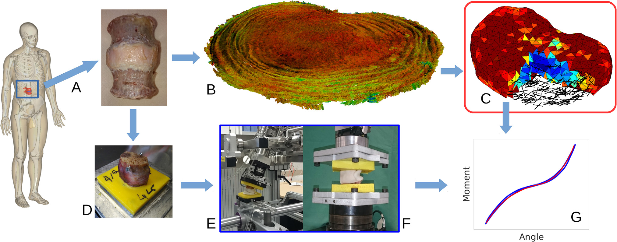
Fig. 1.

Study overview: A) A human L2-L3 functional spinal unit was prepared and scanned in a 9.4 T MR scanner. Proton-density weighted and B) diffusion tensor images were acquired and processed to generate C) a fully MRI-based finite element model (geometry, composition and anisotropy). D) The sample was embedded and in vitro mechanical tests in E) lateral bending, axial rotation, flexion, extension and F) tension and compression were conducted on the sample. G) Using the acquired load-displacement curves, the material parameters were calibrated and the performance of the model was evaluated.
