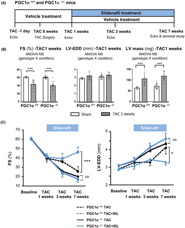
Fig. 1.
Study protocol and echocardiography. (A) A schematic diagram of the study protocol. (B) Fractional shortening (FS), left ventricular (LV) end-diastolic dimension (LV-EDD) and LV mass from echocardiograms of PGC1α+/+ and PGC1α−/− hearts after 1 week exposure to pressure overload (TAC) before sildenafil treatment (n = 14–15 per group). Groups were compared by 2-way ANOVA followed by Tukey-Kramer post-hoc test. (C) Serial echocardiographic assessments of FS and LV-EDD during sildenafil treatment (n = 7–9 per group). Sildenafil treatment (blue) significantly improved FS and ameliorated LV-EDD enlargement in PGC1α+/+ hearts (dotted lines), but not in PGC1α−/− hearts (solid lines). Groups were compared by 1-way ANOVA followed by Tukey-Kramer post-hoc test. *P < 0.05, ***P < 0.001.

