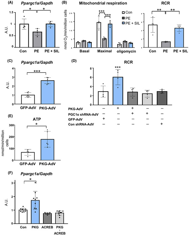
Fig. 4.
cGMP-PKG regulation of mitochondrial respiratory function via PGC1α in rat neonatal cardiac myocytes. (A) PGC1α expression and (B) mitochondrial respiratory function (glutamate/malate substrate) with or without sildenafil (SIL) in RNCMs exposed to prolonged phenylephrine (PE) (n = 4 per group). Groups were compared by 1- or 2-way ANOVA followed by Tukey-Kramer post-hoc test. (C) PGC1α (Ppargc1a) mRNA up-regulation by adenoviral PKGI α induction (n = 6 per group). Groups were compared by unpaired Student’s t test. (D) PKGIα regulation of mitochondrial respiratory function (RCR, respiratory control ratio with glutamate/malate substrate with or without PGC1α silencing) (n = 4 per group). Groups were compared by 1-way ANOVA followed by Tukey-Kramer post-hoc test. (E) ATP generation during state 3 respiration by PKGIα induction (n = 4-5 per group). Groups were compared by unpaired Student’s t test. (F) PGC1α expression by PKGIα in HEK cells with or without ACREB (CREB inhibitor) (n = 6–9 per group). Groups were compared by 1-way ANOVA followed by Tukey-Kramer post-hoc test. *P < 0.05; **P < 0.01; ***P < 0.001.

