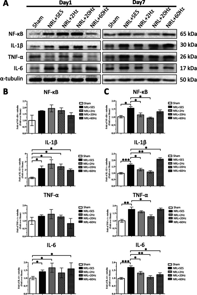
Fig. 4.
Effect of SNS on the expression of spinal cord NF-κB, TNF-α, IL-1β, and IL-6 in L5 NRL rats. Spinal cord tissues were obtained from NRL rats (N = 5) on PID1 and PID7. Tissue lysates were analyzed by immunoblotting with specific antibodies against NF-κB, TNF-α, IL-1β, and IL-6 (a). α-tubulin was used as an internal control. Relative levels of spinal cord NF-κB, TNF-α, IL-1β, and IL-6 on PID1 were quantified by densitometric analysis using ImageJ software (b). Relative levels of spinal cord NF-κB, TNF-α, IL-1β, and IL-6 on PID7 were quantified by densitometric analysis using ImageJ software (c). Data are expressed as mean ± SD. *p < 0.05, **p < 0.01, ***p < 0.001

