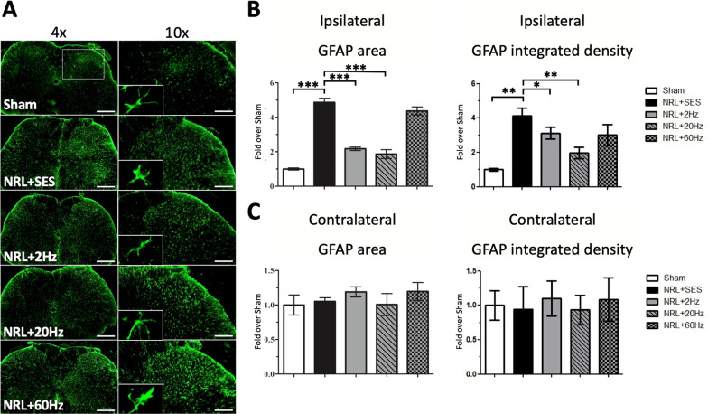
Fig. 5.
Proliferation of SCDH astrocytes on PID7 following SNS at various frequencies in L5 NRL rats. Transverse sections of L5 spinal cord were obtained from NRL rats on PID7 (N = 5). Immunofluorescence staining of the astrocyte marker GFAP (green) on PID7 (a). The right panels show magnification (× 10) of the ipsilateral SCDH. The relative area and integrated density of ipsilateral and contralateral GFAP signal was quantified using ImageJ software (b, c). Scale bars = 500 μm and 200 μm (magnification). Data are expressed as mean ± SD. *p < 0.05, **p < 0.01. ***p < 0.001

