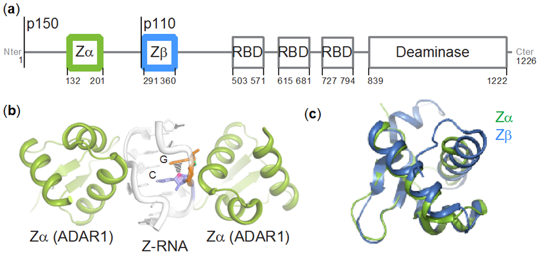
Figure 1. The Zα and Zβ domains of ADAR1p150.

(a) Domain organization of ADAR1: Zα and Zβ are structurally homologous helix-turn-helix DNA-binding domains, RBD stands for double-stranded RNA binding domain. Both isoforms are indicated. (b) Crystal structure of (CpG)3 RNA bound to Zα from ADAR1 (PDB ID: 2GXB, (Placido et al. 2007)). (c) Structural alignment of the Zα (PDB ID: 2GXB) and Zβ (PDB ID: 1XMK, (Athanasiadis et al. 2018)) domains of ADAR1. The backbone RMSD between the two structures is 0.9 Å (excluding the termini).
