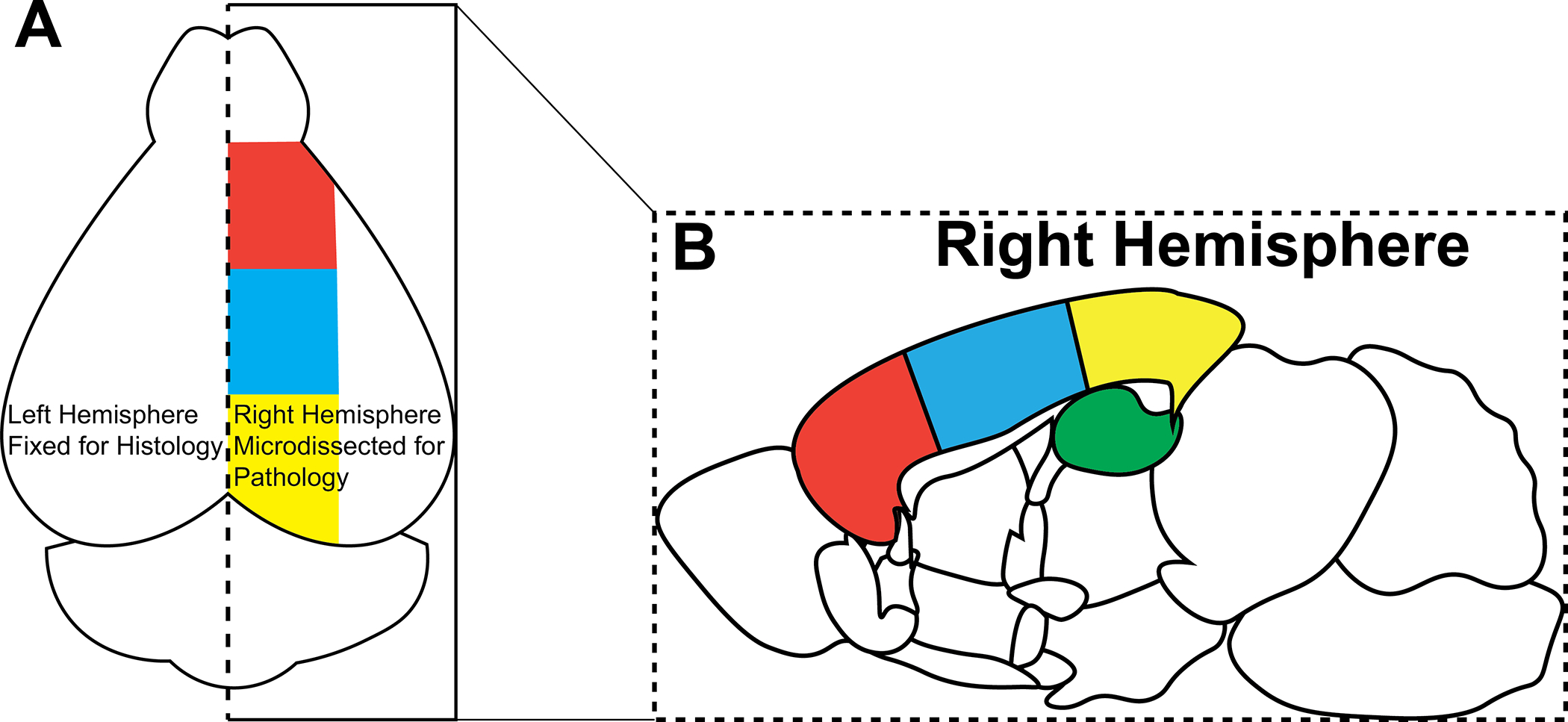
Figure 5: Mouse brain microdissection.

(A) After the brain is extracted from mouse, it is bisected the along dashed line. The left hemisphere is fixed for histology, and the right hemisphere is microdissected for pathology. (B) Sagittal view of the cortex of the right hemisphere. The right hemisphere is microdissected into corresponding color-coded regions. For analysis of freeze-sensitive proteins, it is optimal to sub-divide tissue sections prior to flash freezing.
