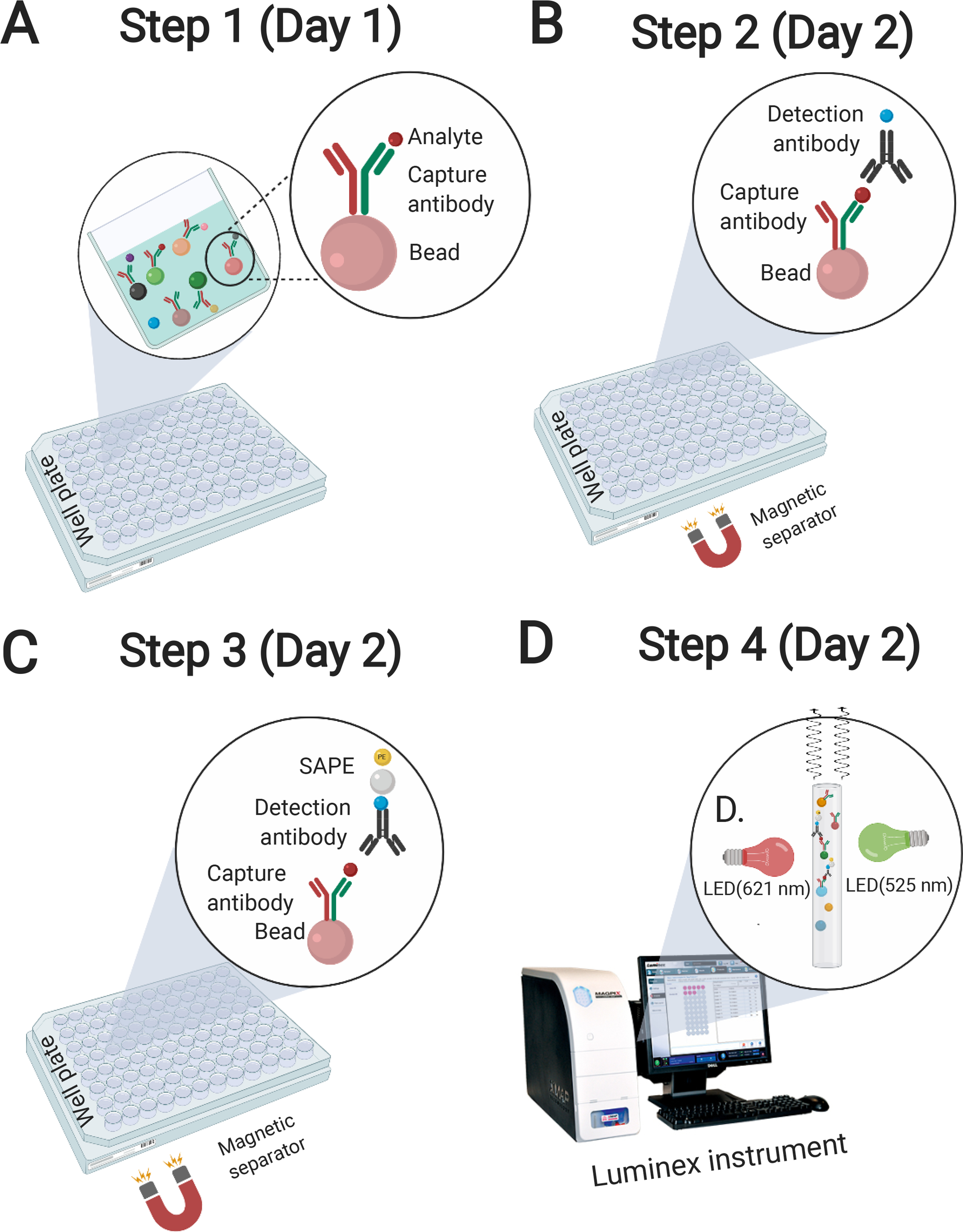
Figure 6. Illustration of Luminex procedure.

(A) Add samples to fluorescently tagged beads. Beads are pre-coated with a specific capture antibody for each protein of interest. (B) Add biotinylated detection antibodies. Biotin-detection antibodies bind to the analytes of interest and form an antibody-antigen sandwich. (C) Add phycoerythrin (PE)-conjugated streptavidin (SAPE). SAPE binds to the biotinylated detection antibodies, completing the reaction. For phospho-proteins, an amplification buffer (only for phospho-protein assays) is added following the addition to SAPE to enhance the assay signal. (D) Luminex instrument (MAGPIX, 200, or FlexMap 3D) reads reaction on each fluorescently tagged bead via a combination of red/green illumination.
