Figure 5.
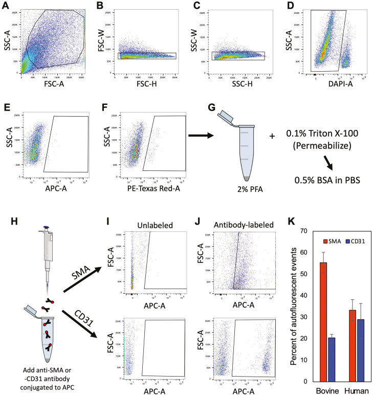
Flow cytometric detection, isolation, and characterization of autofluorescent events in adult cow and human ovarian cortical tissue. (A–D) Representative gating strategy for doublet discrimination (forward-scatter or FSC-A: B; side-scatter or SSC-A: C) and for dead cell exclusion using 4ʹ,6-diamidino-2-phenylindole (DAPI) labeling (D). (E, F) Comparison of autofluorescent events detected in the APC-A far-red channel (640-nm laser; E) versus the PE-Texas red-A channel (561-nm laser; F). (G–K): Autofluorescent events detected in the PE-Texas red-A channel were collected, fixed and permeabilized (G and H), and then incubated with APC-conjugated primary antibodies against SMA (Abcam ab5694) or CD31 (Invitrogen MA3100) (I and J) for determination of the total percentage of autofluorescent events that were positive for expression of either PVC marker in bovine and human ovarian cortical tissue samples (K). Data shown in (K) are the mean ±SE; n = 4 (CD31) or 7 (SMA), and n = 5 (SMA) or 6 (CD31), for bovine and human sample analysis, respectively.

