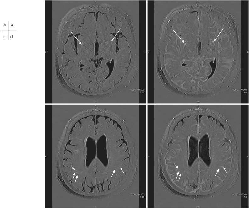
Fig. 1.
A 72-year-old woman with a suspicion of endolymphatic hydrops. 3D-real inversion recovery (TR 15130/TE 549/TI 2700) images of the whole brain were obtained before (a, c) and 4 hrs after (b, d) IV-GBCA for the evaluation of endolymphatic hydrops. A slice at the basal ganglia level (a, b) and a slice at the body of the lateral ventricles level are shown (c, d). The PVS in the basal ganglia shows lower to similar signal intensity compared to the brain parenchyma (a, arrows) in the pre-contrast scan. The PVS in the basal ganglia show marked enhancement at 4 hrs after IV-GBCA (b, arrows). Note that the CSF in the subarachnoid space also shows contrast enhancement (b). The PVS in the white matter shows high signal intensity in the pre-contrast scan (c, short arrows) and no apparent enhancement is seen at 4 hrs after the IV-GBCA (d, short arrows). CSF, cerebrospinal fluid; IV-GBCA, intravenous administration of a single dose of GBCA; PVS, perivascular space.

