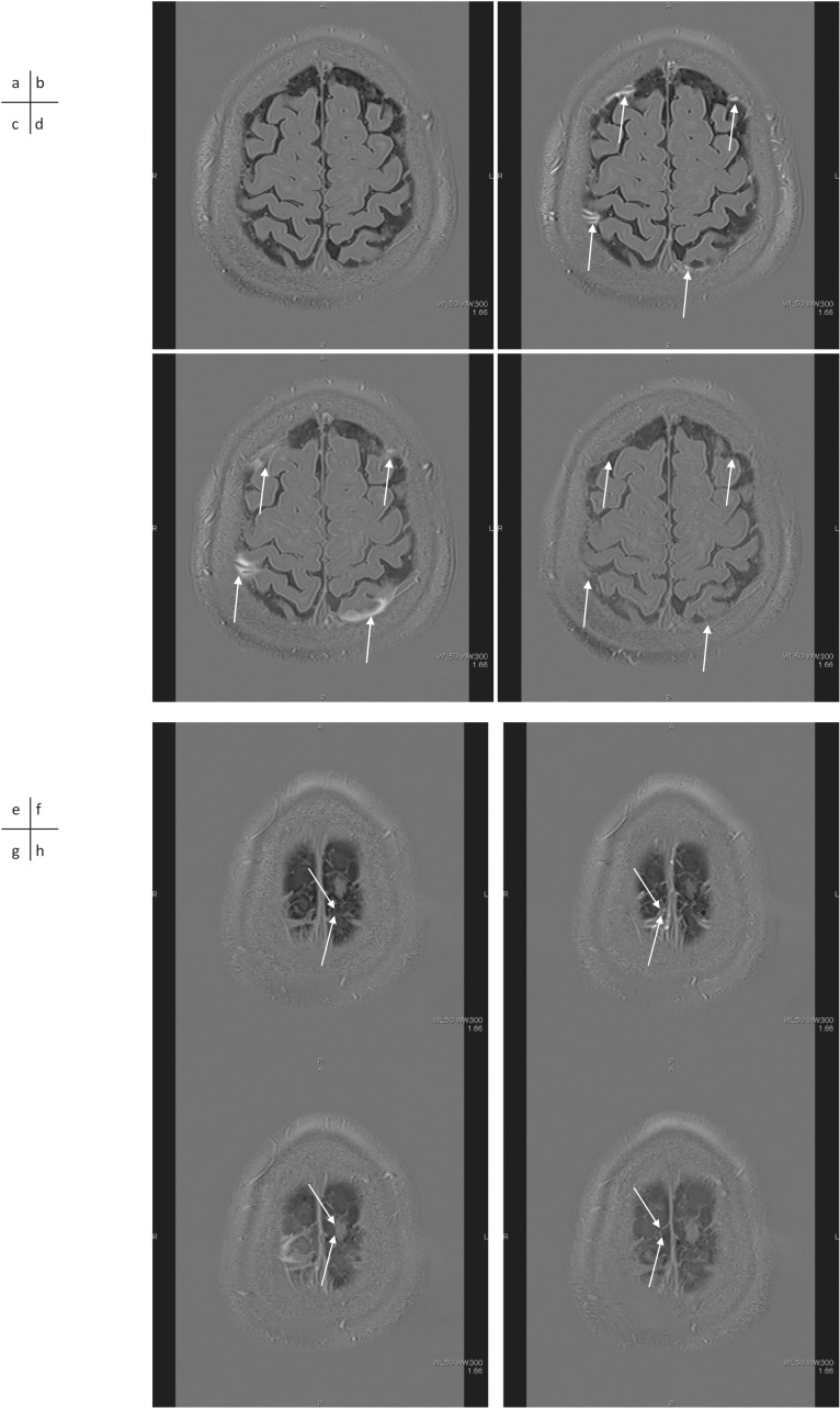
Fig. 2.
A 61-year-old man with a suspicion of endolymphatic hydrops. 3D-real inversion recovery (TR 15130/TE 549/TI 2700) images of the whole brain were obtained before (a), 5 mins (b), 4 hrs (c) and 24 hrs (d) after the IV-GBCA for the evaluation of endolymphatic hydrops. Perivenous enhancement appears at 5 mins after the IV-GBCA (b, arrows) and the enhancement spreads into the surrounding CSF space (c, arrows). The enhancement in the CSF space is markedly decreased at 24 hrs after the IV-GBCA (d, arrows). 3D-real IR images obtained before (e), 5 mins (f), 4 hrs (g) and 24 hrs (h) after the IV-GBCA at a more superior level are shown. A perivenous cyst (arrows) is visualized near the superior sagittal sinus. This cyst might be blocking the flow of the interstitial fluid in the peri-venous subpial space, or the cyst might have formed due to an obstruction of the subpial fluid flow by an unknown cause. CSF, cerebrospinal fluid; IV-GBCA, intravenous administration of a single dose of GBCA.

