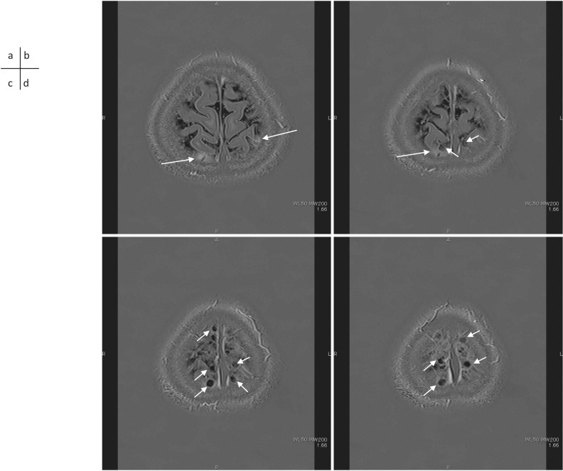
Fig. 3.
A 41-year-old woman with a suspicion of endolymphatic hydrops. 3D-real inversion images and recovery images (TR 15130/TE 549/TI 2700) obtained 4 hrs after an IV-GBCA. The enhancement by GBCA leakage spread into the surrounding CSF space (a, b, arrows). Perivenous cysts (b, c, d, short arrows) are visualized near the superior sagittal sinus. These perivenous cysts are located along the superior sagittal sinus. CSF, cerebrospinal fluid; IV-GBCA, intravenous administration of a single dose of GBCA.

