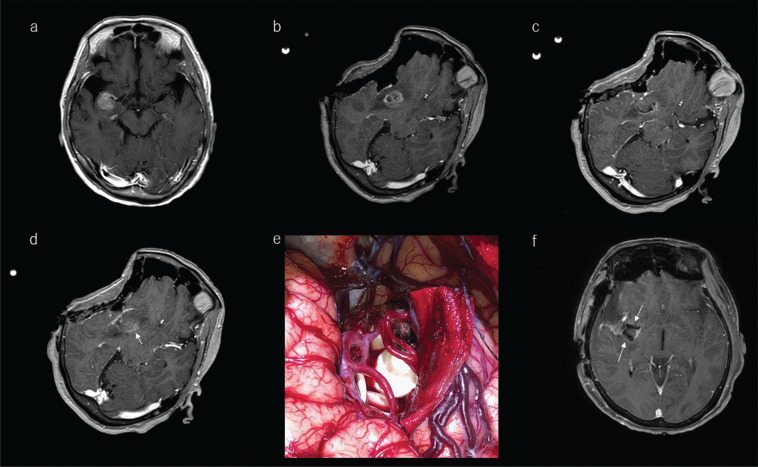
Fig. 10.
Illustrative case of a 72-year-old woman with high-grade glioma treated intraoperatively with a carmustine-impregnated wafer implanted into the tumor cavity. a: Preoperative T1-weighted gadolinium-enhanced MRI showed a lesion located at the anterior part of the right insula. b: After craniotomy, marked brain shift was noted related to drainage of cerebrospinal fluid from the Sylvian fissure. The surgeon decided to use these ioMR images as reference images in the neuronavigation system. c: The ioMR image seemed to indicate the removal of the enhanced lesion. d: One slice above the ioMR image in c showed a small volume of enhanced lesion (arrow). The surgeons decided to leave this remnant in place because it has crossed the pyramidal tract, and to treat it with adjuvant therapy. e: Biodegradable carmustine-impregnated wafers (white materials) were placed in the tumor cavity. f: The carmustine-impregnated wafers appeared with low-intensity signal on the postoperative MR image (arrows). ioMR, intraoperative magnetic resonance.

