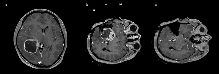
Fig. 2.
Illustrative case of a right-temporal deep-seated glioma showing countermeasures against brain shift. a: Preoperative enhanced T1-weighted images show a ring-enhanced mass lesion with peritumoral edema. In this case, brain bulging after the craniotomy and brain shift are expected to occur during the surgery. b: The surgeon has uncapped the brain outside the lesion and immediately takes reference ioMR images for neuronavigation before brain shift occurs. c: The ioMR images after glioma removal show a nearly total removal. ioMR, intraoperative magnetic resonance.

