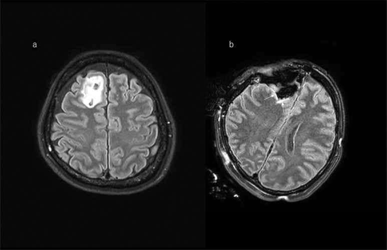
Fig. 7.
Illustrative case of a right frontal glioma. a: A 47-year-old woman with high signal intensity the right frontal lobe on pre-operative FLAIR image. b: Usually ioMR FLAIR images show a linear (like a border of the margin) high signal around the cavity; this should not be misdiagnosed as tumor remnant. FLAIR, fluid-attenuated inversion recovery; ioMR, intraoperative magnetic resonance.

