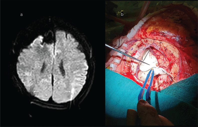
Fig. 8.
Illustrative case of a right frontal glioma. a: A 47-year-old woman with the right frontal lobe glioma. Diffusion-weighted ioMR imaging shows a minimal susceptibility artifact around the cavity. Note also that some artifacts related to the head pin were identified in both occipital lobes (arrows). b: After removing the tumor, the surgeon decides to take intraoperative MR images. Before moving the patient into the intraoperative MR scanner, large enough surgical gauze (with X-ray-enhanced fiber containing polypropylene, barium sulphate, and polyester, which does not affect MR images) is placed into the tumor-removed cavity. It is filled with fluid so that it will not collapse the cavity, and this step prevents the cavity wall falling inward. The cavity is filled with irrigation fluid preventing air bubbles, which can induce susceptibility artifacts on ioMR images. ioMR, intraoperative magnetic resonance.

