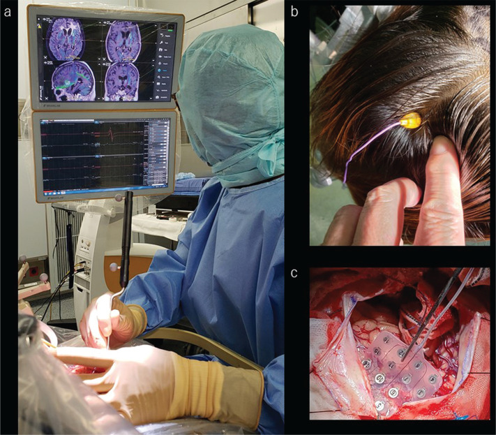
Fig. 9.
Neuronavigation, motor-evoked potentials, and somatosensory evoked potentials provide real-time anatomical and neurophysiological information to the surgeons. a: The surgeon is handling a pointer device and touches the surgical field; the navigation monitor shows the exact position on the upper monitor. The lower monitor shows the motor-evoked potential. b: The evoked potential electrode is screwed into the scalp for transcranial motor-evoked potential monitoring. c: The monitoring electrode is slipped underneath the dura mater for testing of somatosensory and motor-evoked potentials through the cortical surface.

