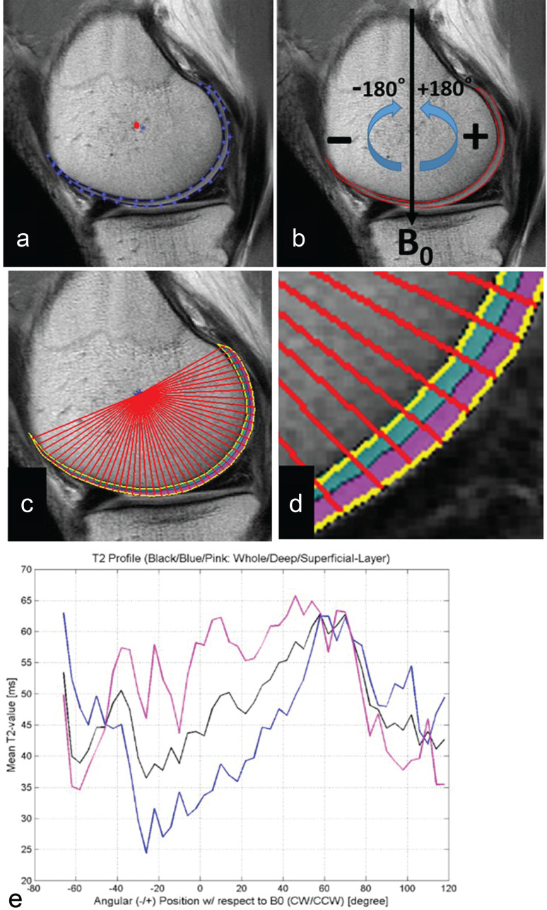
Fig. 2.
Articular segmentation with angle- or layer-dependent approach. (a) After manual cartilage extraction, the central point of the cartilage (red dot) was automatically approximated. (b) Static magnetic field (B0) was defined as 0 degrees, with negative and positive angles located anterior and posterior to the central point. (c) Radial lines from a central point divided cartilage into 4-degree segments. (d) Segmentation of cartilage into deep (0%–50%) and superficial layers (51%–100%) of relative thickness. (e) T2 profiles were generated for whole thickness, deep, and superficial layers of cartilage. (Reprinted with permission from #6).

