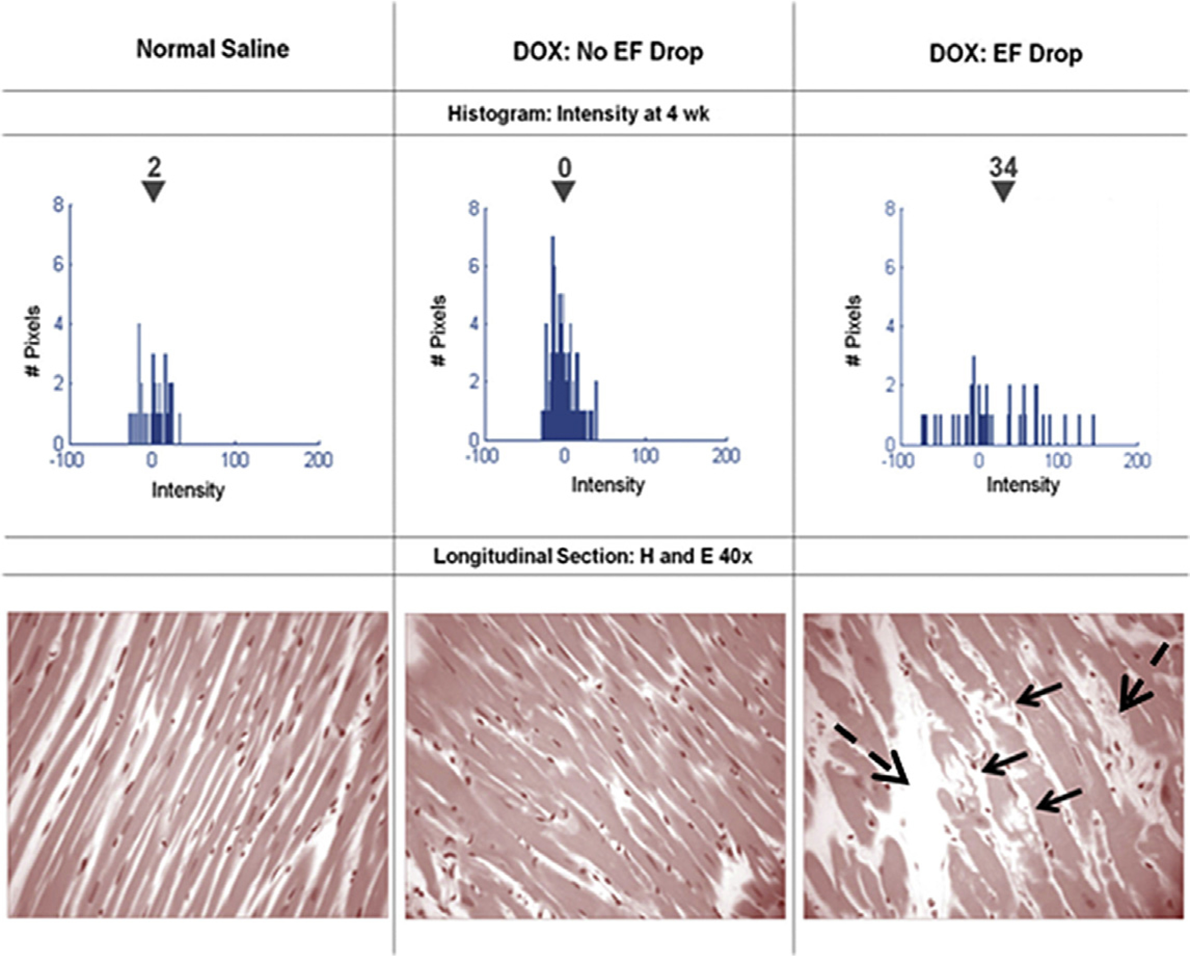
Fig. 7.

Serial histograms of myocardial LGE signal intensity (top, mean intensity shown above the inverted black triangles) and corresponding histopathology (bottom) of individual animals 4 weeks after receipt of normal saline (left), doxorubicin without an LVEF drop (middle), and doxorubicin with an LVEF drop (right). Vacuolization (arrows) and increased extracellular space (dashed arrows) were observed in animals with doxorubicin cardiotoxicity. (From Lightfoot JC, D’Agostino RB, Jr., Hamilton CA, et al. Novel approach to early detection of doxorubicin cardiotoxicity by gadolinium-enhanced cardiovascular magnetic resonance imaging in an experimental model. Circ Cardiovasc Imaging. 2010;3(5):550–558; with permission.)
