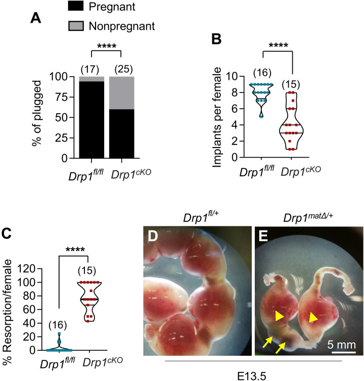
Fig. 3. Postimplantation death of Drp1matΔ/+ embryos derived from Drp1Δ/Δ oocytes.
Five-week-old Drp1cKO and control Drp1fl/fl females were mated with WT (+/+) males and assessed fetal development between E10.5 to E18.5 of pregnancy. (A) Uteri of 94% of plugged control Drp1fl/fl females showed embryos as compared to only 60% of plugged Drp1cKO females contained any embryo. Number of mice analyzed are shown. (B) Control females had an average of eight implants per pregnancy, whereas Drp1cKO females had only four implants per pregnancy. Number of mice analyzed are shown. (C) Within confirmed pregnancies, there was a higher percentage of embryo resorption in Drp1cKO females (76%) compared to only 3% in Drp1fl/fl females. Number of mice analyzed are shown. While the uteri of E13.5 Drp1fl/fl females showed normal developing embryos (D), the embryos in uteri of Drp1cKO females were either dead (E, arrows) or severely growth retarded (E, arrowheads). Results show means ± SD (B and C). P value determined by chi-square test (A), two-tailed Student’s t test (B and C).

