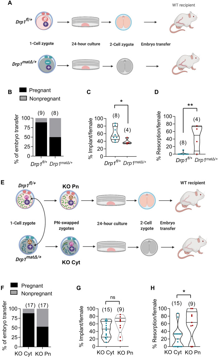
Fig. 6. Development after embryo transfer.
(A to D) Embryonic lethality is due to defective Drp1Δ/Δ oocytes. (A) Schematic representation of embryo transfer experiment. Superovulated 5-week-old Drp1fl/fl and Drp1CKO females were mated with WT (+/+) males to generate WT Drp1fl/+embryos and heterozygous Drp1matΔ/+ zygotes, respectively. One-cell zygotes harvested from the oviducts of plugged donor females were cultured in vitro for 24 hours until they reached the two-cell stage. Two-cell stage embryos were transferred to the oviducts of different WT pseudo-pregnant recipients. (B) Recipients culled between E15.5 and E20.5 of pregnancy revealed that eight of nine recipients receiving Drp1fl/+ embryos became pregnant compared to only four of eight recipients receiving Drp1Drp1matΔ/+ embryos. (C) Per-pregnancy implantation rate of Drp1matΔ/+ embryos was markedly reduced than Drp1fl/+ embryos. (D) Within confirmed pregnancies, there was a higher percentage of embryo resorption in Drp1cKO females compared to Drp1fl/fl females. (E to H) Embryo development after pronuclear swapping and embryo transfer. (E) Schematic representation of reciprocal pronuclear swapping between one-cell zygotes, in vitro culture, and embryo transfer. (F) Comparison of pregnancy rates after implanting KO Cyt and KO Pn zygotes. (G) Implantation rates of KO Pn and KO Cyt embryos. Within positive pregnancies, there was a higher percentage of resorption of KO Pn embryos compared to KO Cyt embryos (H). P values determined by chi-square test (B and F) and two-tailed Student’s t test (C, D, G, and H). Results show means ± SD (C, D, G, and H). Number of mice analyzed are shown (B to D and F to H).

