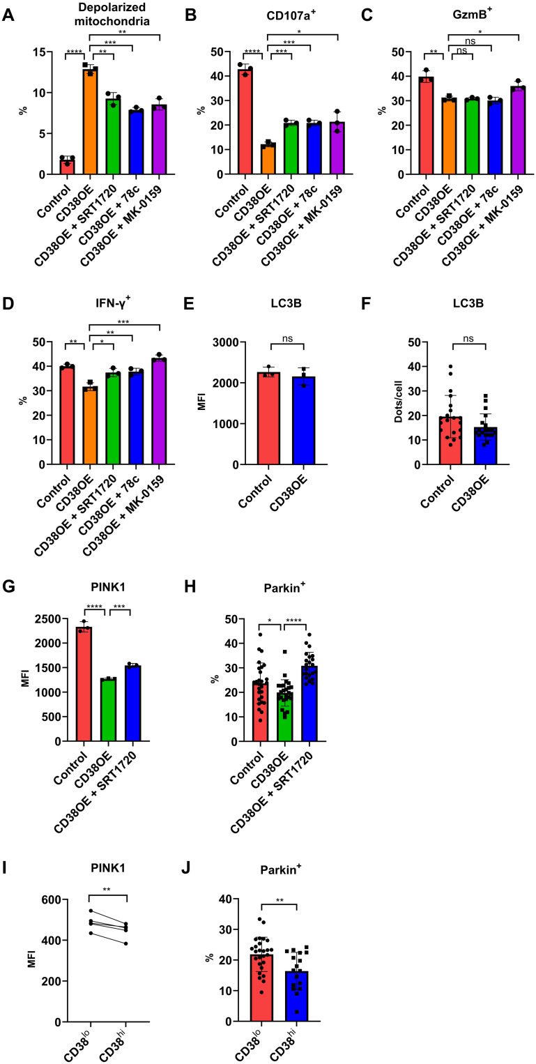
Fig. 2. CD38 perturbs mitochondrial turnover by inhibiting PINK1-Parkin–dependent macroautophagy.
(A) Percentage of depolarized mitochondria in control, CD38-overexpressing CD8+ T cells, or CD38-overexpressing cells treated with SRT1720, or one of the CD38 inhibitors (78c or MK-0159). (B to D) Percentage of positive cells expressing CD107a (B), granzyme B (C), and IFN-γ (D) in control, CD38-overexpressing CD8+ T cells, or CD38-overexpressing cells treated with SRT1720, or one of the CD38 inhibitors (78c or MK-0159). (E) MFI of LC3B staining in control or CD38-overexpressed CD8+ T cells. (F) Counts of LC3B-positive dots per cell of control or CD38-overexpressed CD8+ T cells. (G) MFI of PINK1 in control, CD38-overexpressing CD8+ T cells, or CD38-overexpressing cells treated with SRT1720. (H) Percentage of Parkin positive of the TOMM20-positive area in control, CD38-overexpressing CD8+ T cells, or CD38-overexpressing cells treated with SRT1720. (I) MFI of PINK1 in CD38hiCD8+ and CD38loCD8+ T cells from lupus patient peripheral blood. (J) Percentage of Parkin positive of the TOMM20-positive area in CD38hiCD8+ and CD38loCD8+ T cells from lupus patient peripheral blood. Data are means ± SD; statistical analysis by two-tailed t test (A to H and J), paired t test (I), ns = P > 0.05, *P < 0.05, **P < 0.01, ***P < 0.001, ****P < 0.0001.

