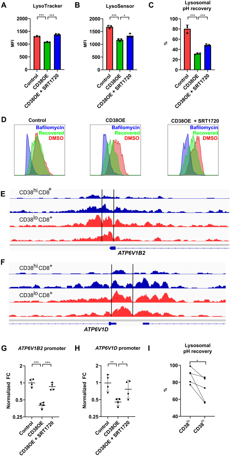
Fig. 3. CD38 regulates lysosomal acidification through epigenetic control of V-ATPase expression.
(A and B) MFI of LysoTracker Deep Red (A) and LysoSensor Green (B) staining in control, CD38-overexpressing CD8+ T cells with and without SRT1720. (C) Percentage of lysosomal pH recovery after removing the reversible V-ATPase inhibitor bafilomycin. (D) Representative flow cytometry plot of LysoTracker Deep Red staining in lysosomal pH recovery test of control, CD38-overexpressing CD8+ T cells with and without SRT1720 in DMSO control condition (red), complete V-ATPase inhibition condition with bafilomycin (blue), and removal of bafilomycin for 1 hour to allow recovery of lysosomal pH (green). (E and F) Sequencing tracks of ATAC-seq data over the promoter region of ATP6V1B2 (E) and ATP6V1D (F) of two replicates of CD38hiCD8+ T cells (blue) and CD38loCD8+ T cells (red) from lupus patient peripheral blood. (G and H) Result of ATAC-qPCR showing normalized fold change (FC) of the two peaks located at the promoter region of ATP6V1B2 (G) and ATP6V1D (H) in control, CD38-overexpressing CD8+ T cells with and without SRT1720. (I) Percentage of lysosomal pH recovery after removing the reversible V-ATPase inhibitor bafilomycin in CD38hiCD8+ and CD38loCD8+ T cells from lupus patient peripheral blood. Data are means ± SD; statistical analysis by two-tailed t test (A to C and G and H), paired t test (I), *P < 0.05, **P < 0.01, ***P < 0.001.

