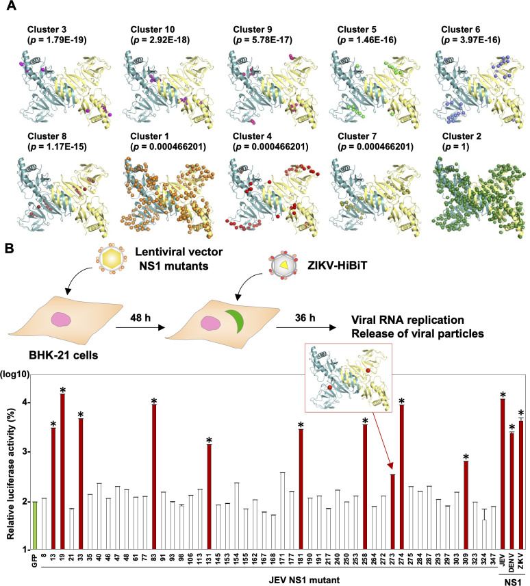
Fig 2. Identification of an amino acid residue necessary for the secretion of NS1 through in silico and mutagenesis analyses.
(A) The protein-protein network within the NS1 protein was analyzed by using co-evolving amino-acid pairs [16, 17] with 66 strains belonging to the Japanese encephalitis serocomplex group. The amino acid residues possessing interaction within clusters are shown as spheres in the three-dimensional structures of NS1 dimers (Protein Data Bank accession number: 4TPL). (B) An illustration shows the experimental workflow. Upon infection with ZIKV-HiBiT at MOI = 0.1, the luciferase activity of cells expressing NS1 mutants carrying the amino acids with different phenotypes (S2 Table) were measured at 36 hpi. The relative luciferase activity was normalized by the activity of cells expressing GFP. The production of double-stranded RNA (dsRNA) was determined by staining with antibody against dsRNA and is indicated by red bars. The locus of amino acid position 273 on the NS1 structure is indicated as red spheres. The data set of the quantified fluorescent signal of dsRNA are found in S2B Fig. Statistical significance was assessed using one-way ANOVA with Dunnett’s test (B) and is indicated by asterisks (*) (versus control cells).

