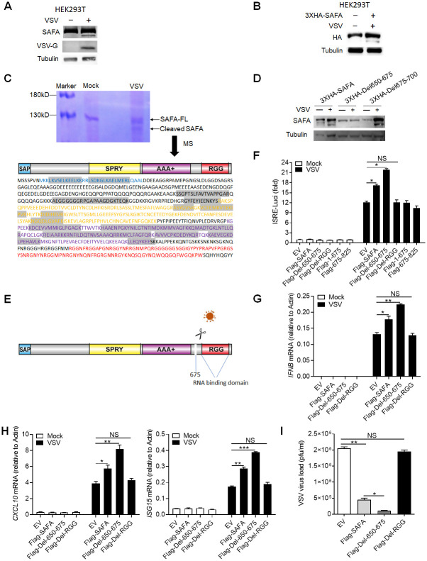
Fig 6. Virus-mediated cleavage of SAFA separates the RNA-binding domain.
(A) Immunoblotting results showing the expression of indicated protein in HEK293T cells infected with VSV for 4 hours. (B) HEK293T cells were transfected with 3XHA-SAFA plasmids, and then infected with VSV for 4 hours followed by immunoblotting. (C) THP-1 cells were infected with VSV for 4 hours followed by immunoprecipitation and coomassie brilliant blue staining. The cleaved band was cut out for mass spectrum assay. The detected amino acid sequences were marked by grey background. (D) HEK293T cells were transfected with indicated plasmids, and then infected with VSV for 4 hours followed by immunoblotting. (E) Models depicting VSV infection induced cleavage of SAFA. (F) HEK293T cells were transfected with indicated plasmids before infection with VSV for 24 hours and then type I interferons in the supernatants were detected by bioassay. (G-I) THP-1 mutants generated by overexpressing indicated lentivirus plasmids were infected with VSV for 24 hours and the expression levels of IFNB (G) and CXCL10, ISG15 (H) were detected by qPCR. The viral load was detected by plaque assay (I). *P < 0.05, **P < 0.01 and ***P < 0.001 (Student’s t-test). The cells were infected by VSV at 0.1 MOI. Data were representative of three independent experiments (A-D). Data were pooled from 3 independent experiments (F-I). Error bars, SEM. n = 3 cultures.

