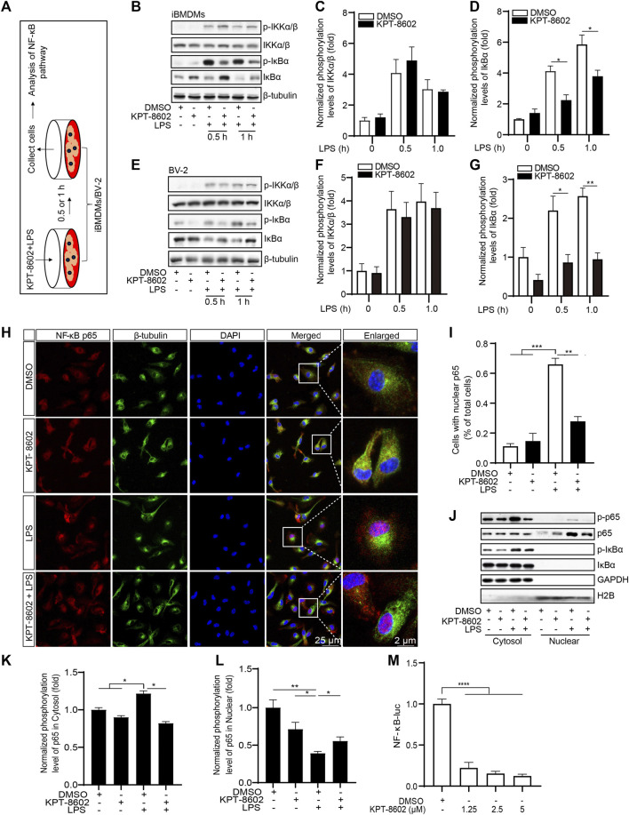
FIGURE 3.
KPT-8602 inhibited LPS-induced activation of the NF-κB signaling pathway. (A) iBMDMs or BV-2 were pretreated with or without KPT-8602 (5 μM) and stimulated with LPS for 0.5 and 1 h, respectively, and then the cell lysates were collected for analysis by Western blotting (B,E). The gray values of the phosphorylated and total IKKα/β (C,F) and IκBα (D,G) bands were analyzed with ImageJ. (H,I) PMs were pretreated with KPT-8602 for 30 min and stimulated with LPS for 2 h. Then, the cells were fixed and stained with a rabbit anti-NF-κB p65 antibody and mouse anti-β-tubulin antibody (H), and the number of cells containing nuclear p65 was analyzed by ImageJ (I). Scale bars, 25 μm for low-magnification images and 2 μm for high-magnification images, respectively. (J,K) iBMDMs were pretreated with KPT-8602 for 1 h and stimulated with LPS for 2 h, and then extracts from the nucleus and cytoplasm were collected for analysis by Western blotting (J). The gray value of the phosphorylated and total p65 in the nucleus (K) and cytoplasm (L) were analyzed with ImageJ. H2B served as a nuclear protein marker; GAPDH as a cytosolic protein marker. (M) Quantitative analysis of the effect of KPT-8602 on NF-κB luciferase activity. (* indicates p < 0.05, ** indicates p < 0.01, *** indicates p < 0.001, **** indicates p < 0.0001 by one-way ANOVA).

