
Figure 1.
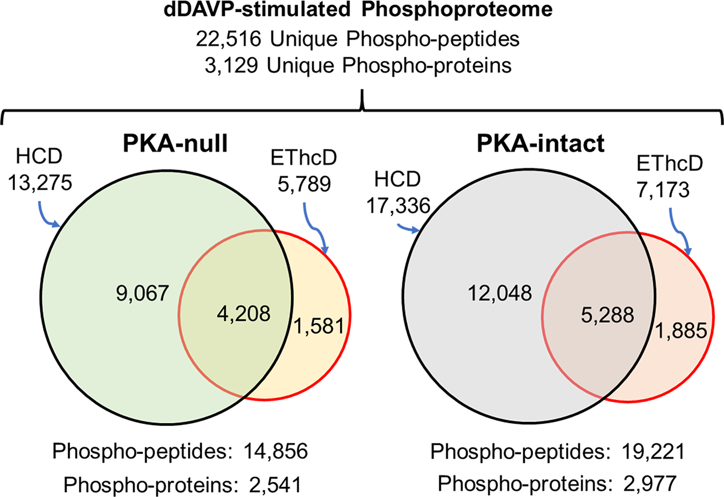
Venn diagrams comparing unique phosphopeptides and phosphoproteins identified by SILAC-based quantitative phosphoproteomics analysis in dDAVP-stimulated PKA-null and -intact cells using HCD and EThcD fragmentation. These phosphopeptides were identified 1) in all three biological replicates for receptive cell-types 2) with a minimum spectral area (MS1 scan) of at least 1.0E7. Peptide FDR was <0.01.