
Figure 7.
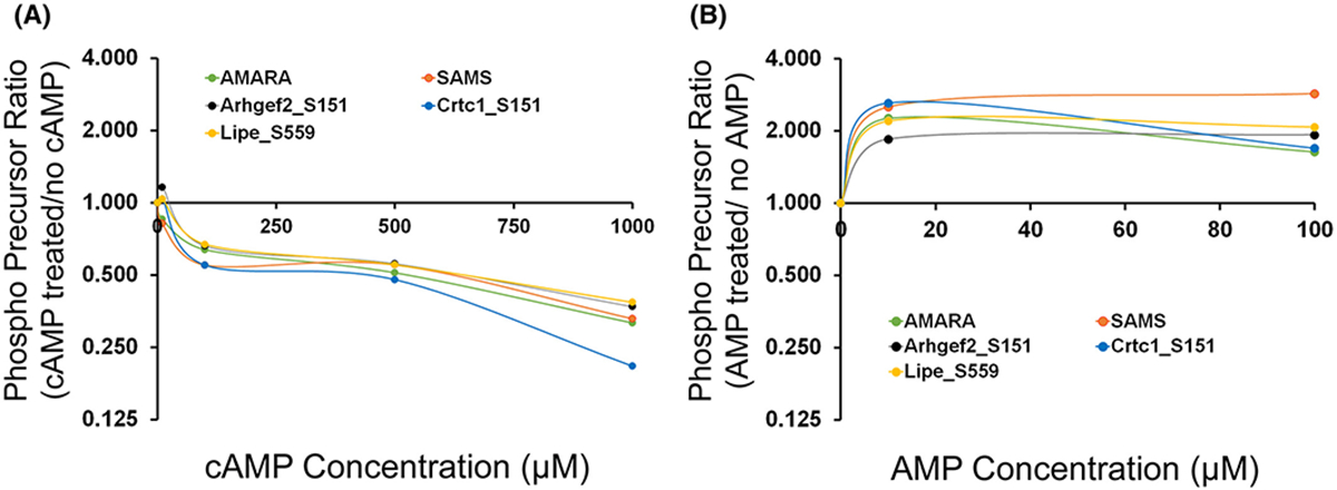
PRM-MS measurement of mono-phosphorylated form of 13-mer synthetic peptides centralized around S151 of Arhgef2, S151 of Crtc1 and S559 of Lipe. Human recombinant AMPK (e.g. AMPKα2β2γ1) that was not phosphorylated at its active site, was used for the in vitro phosphorylation reaction. AMARA and SAMS were used as standard peptide substrates for AMPK phosphorylation. (A) Effect of increasing concentrations of cAMP (0, 10, 100, 500, 1000 μM) on AMPK-induced phosphorylation. (B) Effect of increasing concentrations of AMP (0, 10, 100 μM) on AMPK-induced phosphorylation. X-axis indicates the ratio of chromatographic area of phosphorylated peptides (based on precursor ion chromatogram area) following incubation with various concentrations of (A) cAMP (B) AMP when compared to (A) ‘no cAMP’ or (B) ‘no AMP’ (0 μM). An x-axis value of 1.0 indicates no change.