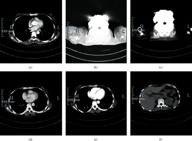
Figure 6.

CT images of one patient. (a)–(c) The CT signs of the patient before treatment. Two small cystic shadows without enhancement are observed in the cervix, about 0.8 cm in diameter. Extensive nodular shadows are observed in the peritoneum, uniform enhancement is observed on enhanced scan, and extensive fluid density shadows are observed in abdominal and pelvic cavities. Extensive fluid accumulation in the abdomen and pelvis. (d)–(f) The CT signs of the patient after treatment. The pelvic mass is significantly reduced, and the pelvic effusion is completely absorbed.
