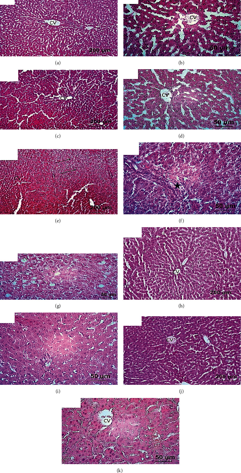
Figure 1.

Light photographs of liver sections stained with H&E. (a, b) Liver sections from the normal rat group showing normal hepatic architecture which comprised of intact hepatic lobules with normal hepatocytes, portal area, and central veins (CV). (c, d) Liver sections from rat received only chrysin (50 mg/kg) showing apparently healthy hepatic lobules with normal hepatocytes appearance. (e, f, g) Liver sections from rats received 20% fructose showing loss of intact liver architecture which characterized by extensive fat droplet disposition (steatosis, dashed arrows), ballooning of hepatocytes and pyknotic nuclei (arrows), and inflammatory cell infiltration (star). Additionally, the blood sinusoids are dilating. (h, i) Liver sections from rat received 20% fructose and concomitantly treated with chrysin (25 mg/kg). (j, k) Liver sections from rat received 20% fructose and concomitantly treated with chrysin (50 mg/kg), showing marked improvement in the hepatic appearance with lesser or completely lacking of fatty disposition and inflammatory cells.
