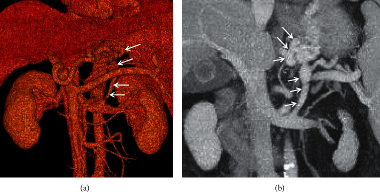
Figure 10.

Gastrorenal shunt. A 62-year-old male with a four-year history of unexplained liver cirrhosis presented with recurrent hematemesis. The contrast-enhanced CT scan on the portal vein phase demonstrated that gastrorenal shunt (white arrow) manifested as enhancement of abnormally dilated vessels originating from the gastric vein to the left renal vein. (a) Three-dimensional reconstruction; (b) coronal contrast-enhanced CT.
