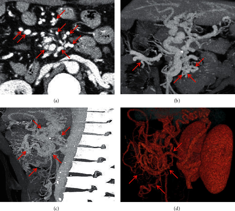
Figure 11.

Retzius vein. A 48-year-old male patient with an eleven-year history of alcoholic-related liver cirrhosis presented with hematochezia. The contrast-enhanced CT scan on the portal vein phase demonstrated that the Retzius vein (red arrow) manifested as tortuously dilated vessels in the retroperitoneum in the portal vein phase. (a) Axial contrast-enhanced CT; (b) coronal contrast-enhanced CT; (c) sagittal contrast-enhanced CT; (d) three-dimensional reconstruction.
