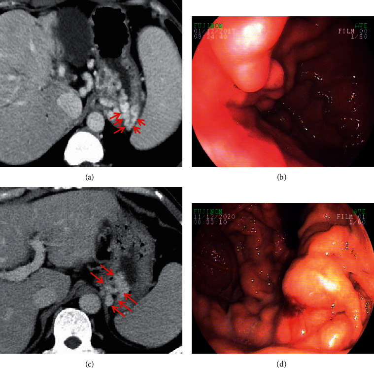
Figure 5.

Gastric varices. In a 47-year-old female and a 56-year-old male with liver cirrhosis, the axial contrast-enhanced CT scan on the portal vein phase demonstrated that gastric varices (red arrow) were long, nodular, and tortuous enhanced channels at the gastric fundus. Upper gastrointestinal endoscopy showed dilated and tortuous varices at the gastric fundus. Note: (a, b) the contrast-enhanced CT scan and endoscopy of a 47-year-old female, respectively; (c, d) the contrast-enhanced CT scan and endoscopy of a 56-year-old male, respectively.
