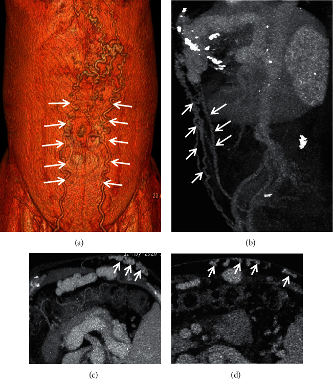
Figure 8.

Abdominal wall varices. In a 65-year-old female with liver cirrhosis, the contrast-enhanced CT scan on the portal vein phase demonstrated that abdominal wall varices (white arrow) manifested as dilated, enhanced, and tortuous structures. (a) Three-dimensional reconstruction; (b) sagittal contrast-enhanced CT; (c, d) axial contrast-enhanced CT.
