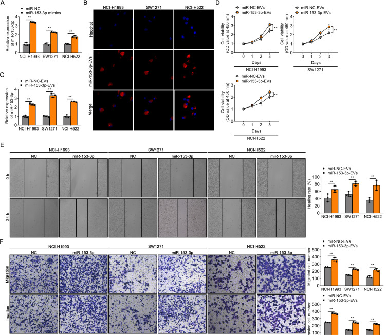
Fig. 3. EV-transferred miR-153-3p enhances the malignant phenotype of LUAD cells.
(A) The expression of miR-153-3p in EVs transfected with miR-NC or miR-153-3p mimics, respectively. (B) Confocal microscopy images of miR-153-3p-EVs uptaking by NCI-H1993, SW1271, and NCI-H522, respectively. MiR-153-3p-EVs were marked using PKH26 and the nuclei were marked using Hoechst. (C) qRT-PCR analysis of miR-153-3p accumulation in LUAD cell lines treated with miR-NC-EVs or miR-153-3p-EVs, respectively. (D) The viability of LUAD cell lines (NCI-H1993, SW1271, and NCI-H522) was assessed at 1, 2, and 3 days after treatment with miR-NC-EVs or miR-153-3p-EVs. (E) Quantification of the wound-healing assay in NCI-H1993, SW1271, and NCI-H522 cells treated with miR-NC-EVs or miR-153-3p-EVs. (F) Quantification of migration and invasion assays in NCI-H1993, SW1271, and NCI-H522 cells treated with miR-NC-EVs or miR-153-3p-EVs. The data in the figures are represented the mean ± SD. **P < 0.01.

