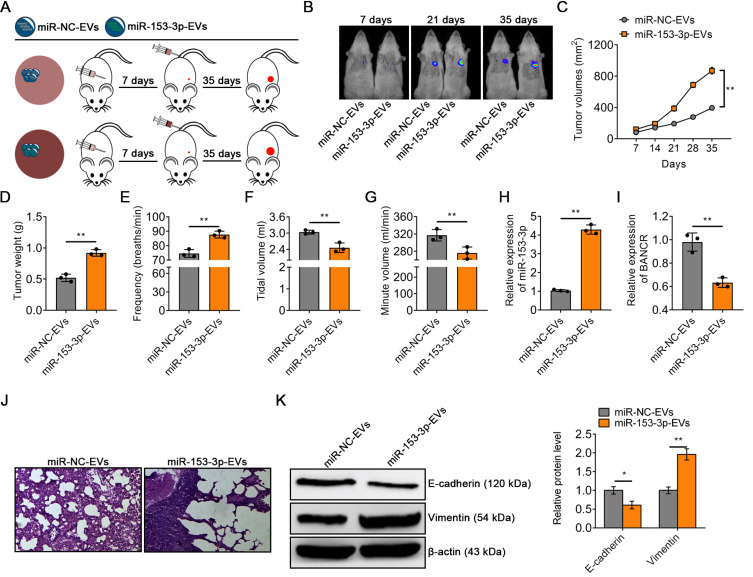
Fig. 5. EVs modified with miR-153-3p mimics contributed to the progression of lung cancer.
(A) Experimental design of mouse model treated with miR-NC-EVs or miR-153-3p-EVs. (B) Representative BLI images of tumor in 6 mice at 7 days, 21 days, and 35 days treated with miR-NC-EVs or miR-153-3p-EVs. (C) Tumor volume was calculated every 7 days after injection. (D) Tumor weight was calculated after tumor excision. Ventilatory function ((E) respiratory rate, (F) tidal volume and (G) minute volume) of mice in miR-NC-EVs group or miR-153-3p-EVs group. (H) qRT-PCR analysis of miR-153-3p expression in the tumor treated with miR-NC-EVs or miR-153-3p-EVs in vivo. (I) qRT-PCR analysis of BANCR expression in the tumor treated with miR-NC-EVs or miR-153-3p-EVs in vivo. (J) H&E staining of lung tissue morphology of mice in the miR-NC-EVs group or miR-153-3p-EVs group. (K) Western blot analysis of E-cadherin and vimentin in tumor in vivo treated with miR-NC-EVs or miR-153-3p-EVs. The data in the figures are represented the mean ± SD. *P < 0.05; **P < 0.01.

