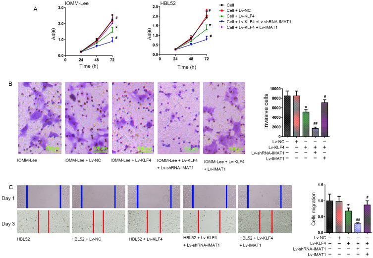
Fig. 4. Effect of overexpression and knockdown of IMAT1 on effects of KLF4 to cellular processes in meningioma cells.
(A) Proliferation of IOMM-Lee and HBL52 cells was determined 72 h after infection with the indicated viruses using CCK-8 assay. (B) Invasion of IOMM-Lee cells was determined using the transwell assay 72 h after infection with the indicated viruses. (C) Migration of HBL52 cells was determined by scratch wound healing assay 72 h after infection with the indicated viruses. Images of cells were used to observe the number of cells growing across the scratched lines. The number of cells growing across the scratched line was defined as the number of migrated cells, and used to compare the changes in the migration ability in each group. *P < 0.05, vs cell group; #P < 0.05, ##P < 0.01, vs cell + Lv-KLF4 group (t-test). The tests were carried out in biological triplicates, and data are expressed as the mean ± SD.

