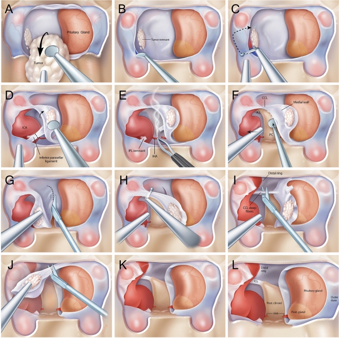
Figure 1.
Medical illustrations demonstrating the sequential steps of resecting the medial wall of the cavernous sinus. Wide exposures of the anterior cavernous sinus and paraclinoid carotid artery allow for adequate visualization and safe removal of the medial wall. (A) Pituitary adenoma is resected under high resolution endoscopy with bimanual technique preserving the normal pituitary gland (B) The medial wall is adequately visualized with high resolution angled endoscopes once the bulk of the adenoma has been removed, tumor remnants that are intraoperatively viewed to adhere strongly to the medial wall suggests invasion necessitating resection. The anterior cavernous sinus is entered by developing a plane between the 2 layers of dura of the intercavernous sinus (C) Using a right angled blunt tipped feather blade, the anterior cavernous sinus wall is cut and hemostasis is achieved (D) The inferior parasellar ligament (IPL) is one of the first ligaments encountered and is cut to allow mobilization of the medial wall away from the carotid artery (E) The inferior hypophyseal artery is often encountered next and is coagulated and cut to avoid any evulsions off the carotid artery (F) the dura overlying the base of the posterior clinoid forms the posterior wall of the cavernous sinus and is dissected off the clinoid till the surgeon encounters the horizontal fibers of the carticoclinoidal ligament (CCL) (G) Microscissors are used to cut the dura covering the dorsum sella to deatch the meial wall from its sellar attachments (H) Using the right angled feather blade, the CCL fibers are cut to begin detaching the medial wall from the carotid artery (I) There are often deep fibers of the CCL that require further transection to completely untether the medial wall from the carotid artery (J) Once all the fibers of the CCL are cut, the medial wall is now completely free from any attachments to the carotid artery and the only remaining cut are dural attachments that make up the proximal dural ring (K) The medial wall is now completely free from attachments and often sent en bloc if it is not severely distorted by tumor invasion leaving behind and open cavernous sinus with an exposed carotid artery. (L) The remaining view should show the medial surface of the carotid artery in the cavernous sinus with visualization of the superior compartment of the cavernous sinus and the interclinoidal ligament (ICL).

