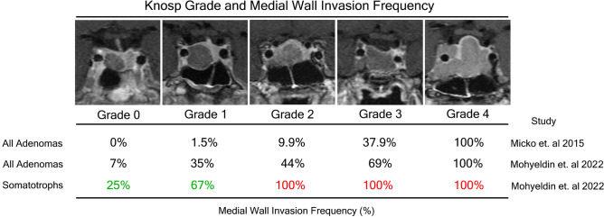
Figure 3.
Knosp grade and medial wall invasion frequency as reported by Knosp and colleagues compared to invasion frequencies exhibited in the current study among all adenomas and then among somatotroph adenomas. Representative MRI images from case examples of somatotrophs in our current series stratified by Knosp grade. Knosp criteria were applied based on carotid tangents and each grade is represented by a coronal T1 MRI scan with gadolinium capturing the extent of cavernous sinus invasion.

