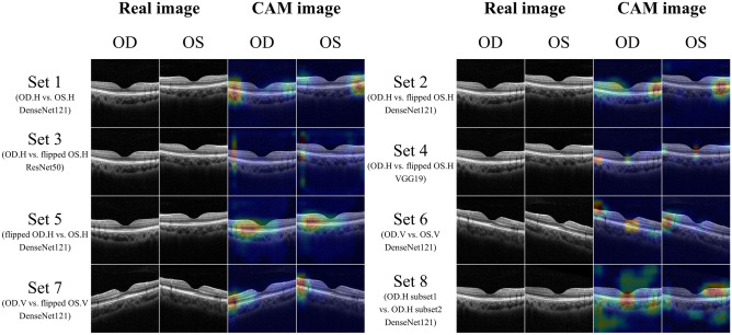
Figure 5.
Image sets and their corresponding class activation mapping (CAM) results. Set 1 consisted of horizontal images of right and left eyes. Papillomacular retinal nerve fiber layers with high reflectivity were shown on the nasal side of the image. CAM highlights both ends of parafoveal regions of images. Sets 2–4 consisted of right horizontal images and flipped left horizontal images. Papillomacular retinal nerve fiber layers with high reflectivity were shown in the right-side of OCT images. CAM patterns in Sets 2–4 differed: DenseNet121 highlighted the parafovea, whereas ResNet50 highlighted only the parafovea and VGG19 both the parafovea and macula. Set 5 consisted of flipped right and left horizontal images; CAM highlighted the parafovea, similar to Set 2. Set 6 consisted of vertical OCT images of right and left eyes. From Set 6, heatmaps highlighting the parafovea and macular region were produced. Set 7 comprised non-flipped right vertical images and flipped left vertical OCT images; the ends of the parafoveal regions were highlighted. Set 8 consisted only of right horizontal images. Unlike Sets 1–7, Set 8 images showed an irregular pattern, emphasizing the vitreous region and outside of the sclera. OD oculus dexter, OS oculus sinister, H horizontal, V vertical.

