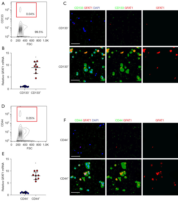
Figure 2.
GFAT1 is highly expressed in CD133+ and CD44+ CSCs in PC. (A-C) In order to examine whether GFAT1 may be associated with PC stemness, CD133+ vs. CD133− fractions were isolated from the PC cell line PANC-1 by flow cytometry. (A) Representative flow chart. (B) RT-qPCR for GFAT1 in the CD133+ population vs. CD133− population. (C) Immunocytochemistry for GFAT1 in the CD133+ population vs. CD133− population from cytospun cells. (D-F) In order to examine whether GFAT1 may be associated with PC stemness, CD44+ vs. CD44− fractions were isolated from the PC cell line PANC-1 by flow cytometry. (D) Representative flow chart. (E) RT-qPCR for GFAT1 in the CD44+ population vs. CD44− population. (F) Immunocytochemistry for GFAT1 in the CD44+ population vs. CD44− population from cytospun cells. DAPI was used for nuclear staining. *, P<0.05. N=5. Scale bars are 50 µm. FSC, forward scatter; GFAT1, glutamine-fructose-6-phosphate transaminase 1; DAPI, 4',6-diamidino-2-phenylindole; CSC, cancer stem cell; PC, pancreatic cancer; RT-qPCR, reverse transcription-quantitative polymerase chain reaction.

