Figure 3.

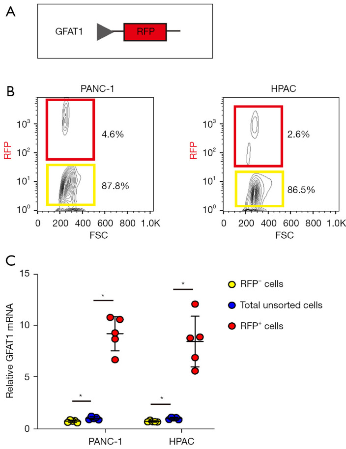
Separation of GFAT1+ vs. GFAT1− PC cells with genetic labeling of RFP. (A) PC cells were transfected with plasmids carrying an RFP reporter under the control of a GFAT1 promoter (pGFAT1-RFP). (B) Two human PC cell lines, PANC-1 and HPAC, were transfected with the designed plasmid. The red fluorescence was analyzed by flow cytometry. Red box indicated GFAT1+ PC cells and yellow box indicated GFAT1− PC cells. (C) RT-qPCR for the mRNA level of GFAT1 in RFP+ and RFP− cells compared to total unsorted cells. *, P<0.05. N=5. RFP, red fluorescent protein; GFAT1, glutamine-fructose-6-phosphate transaminase 1; FSC, forward scatter; PC, pancreatic cancer; RT-qPCR, reverse transcription-quantitative polymerase chain reaction.
