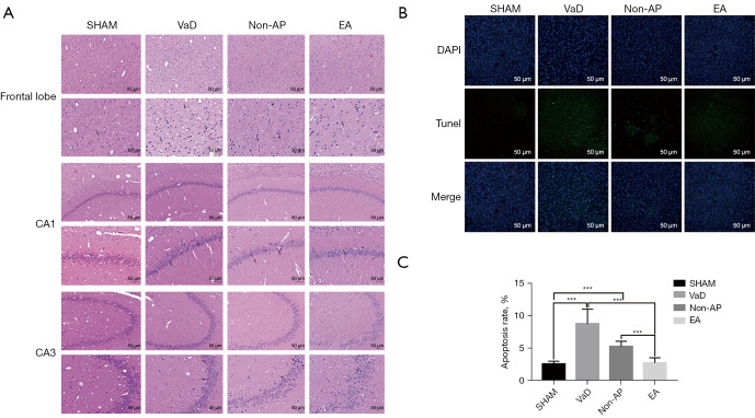
Figure 3.
Histopathological changed in the frontal lobe, and hippocampal CA1 and CA3 regions (n=6/group). (A) Representative images of morphological changed in cerebral cortex, the hippocampal CA1 and CA3 regions in the sham, VaD, non-AP, and EA groups (n=6/group), 2 weeks after EA staining (HE staining; magnification ×200 and ×400). Scale bar, 50 µm. Nuclear pyknosis was indicated with black arrow. (B) TUNEL staining for apoptosis detection. Expression of apoptotic factors in the cortices of rats among all four groups. Scale bar, 50 µm. (C) Apoptosis rate in the cortex of rats. Data were shown as mean ± SD values, and statistical significance between both groups was defined as ***P<0.001. VaD, vascular dementia; AP, acupuncture; EA, electroacupuncture; HE, hematoxylin and eosin; TUNEL, terminal deoxynucleotidyl transferase-mediated dUDP nick-end labeling; CA1, cornu ammonis 1; CA3, cornu ammonis 3.

