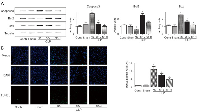
Figure 4.
SF inhibited cardiomyocytes apoptosis in septic mice. (A) Apoptotic protein cleaved caspase3, Bax, and anti-apoptotic protein Bcl2 levels were analyzed by immunoblotting. Data were normalized using tubulin. (B) Representative TUNEL staining of the left-ventricular myocardium section. Blue, DAPI stained nuclei. Original magnification, 400×; scale bar, 100 µm. Quantification of TUNEL positive cells was shown in right panel. &, P<0.05 vs. sham; #, P<0.05 vs. CLP; *, P<0.05 vs. treated with SF-L. SF, Shenfu injection; SF-L, 10 mg/kg SF; SF-H, CLP + 40 mg/kg SF; CLP, cecal ligation and puncture; NS, normal saline; TUNEL, terminal-deoxynucleotidyl transferase mediated nick end labeling.

