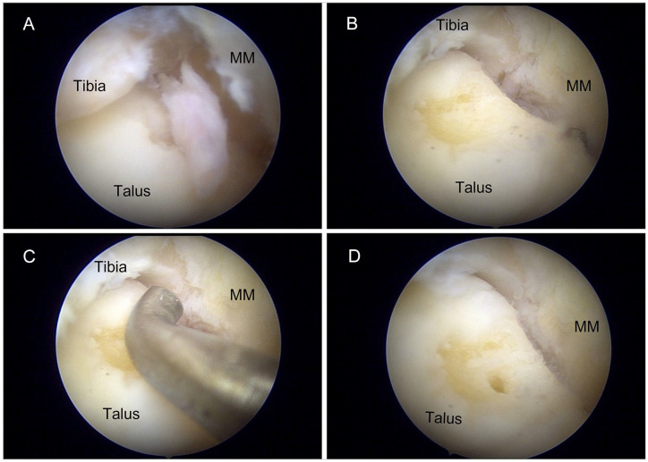
Figure 1.
Intraoperative images of arthroscopic debridement and microfracture of OLTs. (A) OLT was identified by the flap of cartilage overlying the chondral defect. (B) Surrounding loose cartilage was debrided until smooth edges were observed. (C) Size of the lesion was measured with the aid of an arthroscopic probe. (D) Microfracture was performed with an arthroscopic awl to a depth of 2 to 4 mm at 3- to 4-mm intervals until fat globules were observed. MM, medial malleolus; OLT, osteochondral lesion of the talus.

