FIGURE 1.

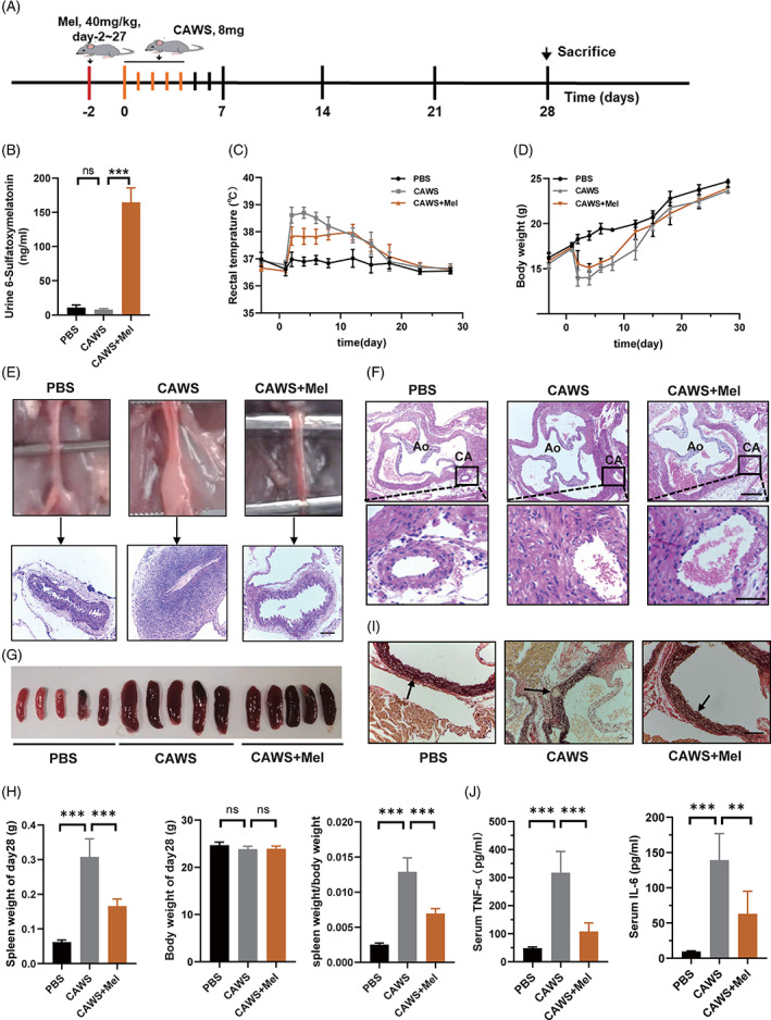
Melatonin alleviates CAWS‐induced KD‐like vasculitis in mice. (A) Modelling timeline of the mouse model. C57BL/6 mice in the CAWS group received i.p. injection of 8 mg CAWS for five consecutive days, while mice in the CAWS+Mel group received i.p. injection of 8 mg CAWS for five consecutive days and that of melatonin (40 mg/kg) for 30 days, and mice in the PBS group received were i.p. injection of 0.1 ml PBS at the same time. (B) Concentration of melatonin metabolite 6‐sulfatoxymelatonin in mice urine, as analysed using ELISA. (C) Mean daily rectal temperature change and (D) mean daily body weight change of mice in the three groups. (E) Histopathological changes in the abdominal aorta were examined using H&E staining, and representative images of the abdominal aorta cut on a transverse plane are shown. Scale bar: 200 μm. (F) Representative H&E images of the aortic root and coronary arteries cut on a transverse plane. Scale bar of the upper pictures is 200 μm and that of the lower pictures is 50 μm. (G) Representative images of the mice spleens on day 28. (H) Spleen weights, body weights, and the ratio of the two on day 28. (I) Representative images of Verhoeff's van Gieson staining of the aortic root on day 28, where arrows indicate the vascular elastic fibre. Scale bar: 100 μm. (J) Serum TNF‐α and IL‐6 concentrations of the mice on day 14, as analysed using ELISA. Data are presented as mean ± SEM of three independent experiments. **p < 0.01, ***p < 0.001; ns, not significant. CA, coronary artery; Ao, aorta; CAWS, Candida albicans water‐soluble fraction; Mel, melatonin; PBS, phosphate‐buffered saline
