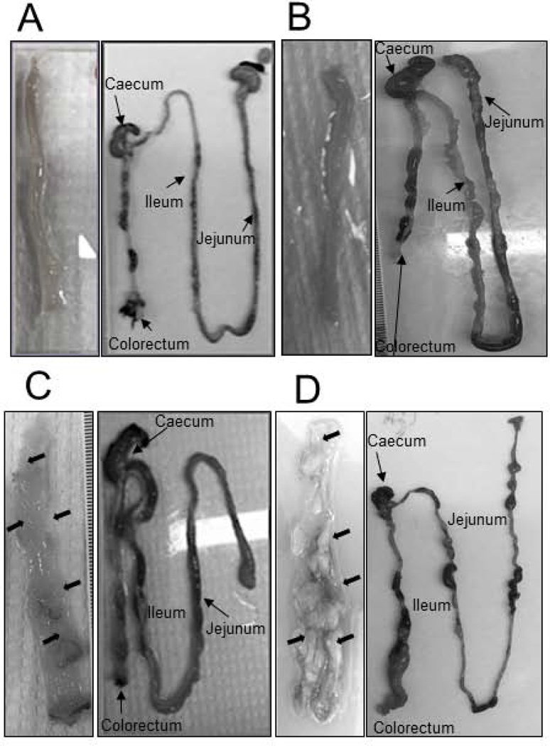
Figure 3.
Macroscopic evaluation of gastrointestinal tissues. Representative images of tissues from ApcW/F/Aldh1b1−/− (A), ApcW/F/Aldh1b1+/+ (B), ApcW/FCdx2ERT2−Cre/Aldh1b1−/− (C) and ApcW/FCdx2ERT2-Cre/Aldh1b1+/+ (D) mice on day 18 of the experimental protocol are shown. Right panels show a low power magnification of the small and large intestine, with jejunum, ileum, caecum and colorectal regions highlighted. Left panels show a higher magnification (A 100X, B 100X, C 400X, D 400X) of the luminal surface of the colon. Macroscopically-observable growths within the colon are indicated by arrows.

