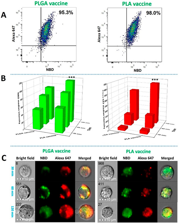
Figure 2.
Cellular uptake and processing of nicotine vaccine particles by DCs. Two million DCs were incubated with 0.5 mg of PLGA nicotine vaccine or PLA nicotine vaccine, of which the lipid layer was labeled with NBD (green) and the KLH was labeled with Alexa 647 (Red), for 30, 60, and 120 min, respectively. The uptake and processing of vaccine particles by the DCs were quantitatively and qualitatively studied using a BD FACSARIA flow cytometer and an Amnis ImageStream flow cytometer, respectively. (A) The percentages of the cells that took up the PLGA and PLA vaccine particles within 120 min. (B) NBD and Alexa 647 median intensities from the cells that were incubated with the vaccine particles for a given period of time. (C) Representative images of processing of vaccine particles in the DCs. The scale bars represent 10 μm. *** denotes that significantly more PLA vaccine was taken up by DCs than PLGA vaccine at 120 min.

