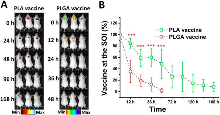
Figure 4.
In vivo degradation of vaccine particles in mice. The protein antigen KLH in the vaccine was labeled with Alexa 750 and 2 mg of the PLGA vaccine, or the PLA vaccine was subcutaneously injected into female BALB/c mice. The degradation of the vaccine particles in the mice was monitored using an IVIS SpectrumCT. (A) The fluorescence image of vaccine particles at the SOI. (B) The relative amount of nondegraded vaccine particles at the SOI. *** denotes that the amount of remaining PLA nicotine vaccine was significantly higher than that of the PLGA nicotine vaccine at the SOI.

